25th March 2024 – by Aaruthy Suthahar
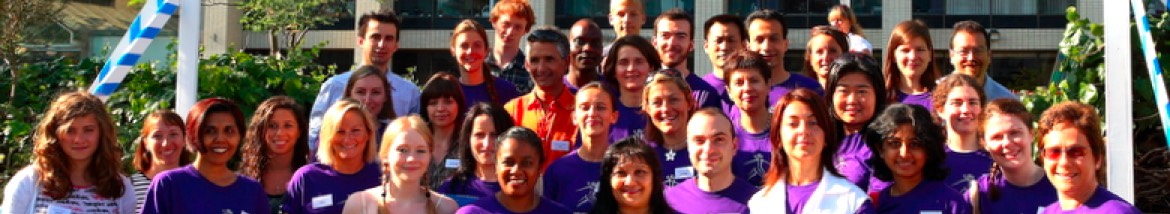
International Women’s Day (IWD) on March 8th is a day to celebrate the achievements and contributions of women globally. At TwinsUK, this year’s IWD was marked with heartfelt stories and reflections from both twins and staff members, showcasing the resilience, strength, and camaraderie that defines the twin community and the workplace culture at TwinsUK.
Twin Stories
Sue, reflected on her late twin sister Jill’s impactful work:
IWD meant nothing to me, but it did to my twin, best known as Jill Saward. Before her death in 2017, she worked tirelessly for women who’d been subjected to Rape or sexual abuse, and victims/survivors of domestic abuse. As identical twins it sometimes made life difficult. People felt through her work they knew me, but also total strangers would ask me how she was. Her impact really hit me after her death. Her friends and campaigners became my friends too – all women.
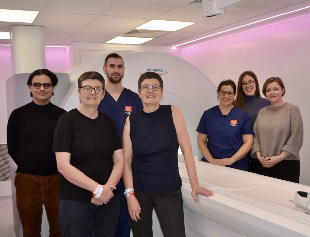
Francina and Vanessa, facing the daunting challenge of breast cancer, found strength in their twin connection.
We are facing one of our biggest challenges this international women’s day year as identical female twins with one of us being diagnosed with breast cancer. Having a mastectomy and facing chemotherapy all I could think about was how different I would look to my twin. We even joked about Vanessa wearing a head scarf when we go out as we love the “ are you twin” comments . We are both clinicians and Christian’s giving us resilience and the ability to face challenges head on. Vanessa has been a constant support and we are celebrating that the genetic test came back negative , reducing my twins risk of having breast cancer. I feel safe knowing my twin is there and am so grateful for my older sister as well. My prognosis is good so looking forward to beating my cancer and getting back to “double trouble“ outings with my twin.
Laura and Jill celebrated not only their twinship but also the joy of raising twin girls themselves:
In honour of International Women’s Day I’m celebrating my amazing identical twin sister Jill who herself has identical twin girls, Aurora ‘Rory’ and Clara – they recently turned 18 months old! They are the coolest and funniest girls on the planet and I watch them with absolute fascination. It is mine and my twin’s duty to set the best example to them of how lucky it is to be a twin and to celebrate it! In the photo, completely unplanned, we are all coordinating tops and bottoms (as are the front doors in the houses behind us).
Margaret and Barbara honored their mother’s legacy, recognising the sacrifices she made in raising them as twins.
I would like to celebrate mothers of twins! Our mother never grew tired of telling the story of how she had the surprise of her life when she was told there’s another baby after I was born. It can’t have been easy bringing up twins in the 1960’s and we only really appreciated this when my son and his fiance became the parents of twins in 2022! Watching them cope with tiny twins night and day has raised so many questions about our own early years. Sadly, we are not able to ask our mum about it as she passed in 2020. The photo attached was taken on her last Mother’s Day, during lockdown when we could not bear to leave her alone all day. Barbara and I will be forever grateful for everything she did for us.
Thoughts from TwinsUK staff
Aaruthy, Communications and Engagement Officer:
Working in the field of science research not only as a woman but also a woman of colour comes with its own set of challenges and opportunities due to lingering stereotypes and biases globally. Yet, at TwinsUK, as someone who is a woman of colour, I’ve had the privilege of experiencing a workplace where diversity and inclusion is embraced. Most of the staff in our department are women from all different backgrounds and each with their own talents and perspectives. I am truly inspired by the women I work with as we collaborate, support each other, and celebrate each other’s successes. I remain hopeful for a future where every woman, regardless of background, can thrive and succeed in the scientific research community.
Bridget, Senior Research Nurse at TwinsUK:
I’ve dedicated the majority of my nursing career to the fields of hematology, oncology, and palliative care, where I’ve been surrounded by compassionate and incredible women. Nursing is undeniably women-centric. My journey led me to TwinsUK, where I gained a glimpse into the world of science, and I’ve cherished every minute of it. Being surrounded by so many intelligent and supportive women from diverse backgrounds has been truly inspiring. Our department is led by fantastic women who wear multiple hats – they’re researchers, clinicians, leaders, and mothers all rolled into one. While there’s still some lingering gender bias in the scientific realm, I remain optimistic that we are making strides in the right direction, one step at a time.
Ayrun, Clinical Operations Manager at TwinsUK:
After my studies, I went from a laboratory assistant to the role of clinical operations manager within my department. Along this journey, I also welcomed two babies, which naturally brought challenges of balancing work with raising young children. However, being part of an inclusive and supportive department where you are made to feel secure makes navigating these challenges easier. What does International Women’s Day mean to me? It means celebrating women’s achievements and contributions- you only need to look at the remarkable women within our department! Equally, it reminds us to recognise the ongoing challenges women still face globally. It is also a day to promote solidarity, to empower, inspire, and uplift women of future generations.
Gulsah, Resource Admin Manager at TwinsUK:
Through my studies of Social sciences from secondary to University years, I was always made hyper-aware of the gender gaps that were statistical facts within our society but admit I had internalised these on some level. As a young, second-generation immigrant female, I did not have the confidence to break these barriers – which was until I started working for the Department of Twin Research. Being surrounded by incredibly successful women from all kinds of backgrounds and walks of life changed my perspective entirely which ignited a new level of ambition in me. As the new Resource Admin Manager, and also a recent first-time mother, I am inspired daily by my remarkable colleagues (both professionally and personally) and couldn’t be prouder to be a woman in science celebrating International Women’s Day here at TwinsUK.
As we reflect on the stories shared by twins and TwinsUK staff, one thing becomes clear: the essence of International Women’s Day extends beyond a single day of celebration. It’s about honouring the past, celebrating the present, and advocating for a future where every woman, regardless of background or circumstance, can thrive. Whether it’s through sisterhood, solidarity, or mentorship, the spirit of IWD lives on in the collective efforts of women everywhere. At TwinsUK, it’s a spirit that continues to inspire and empower each and every day.